
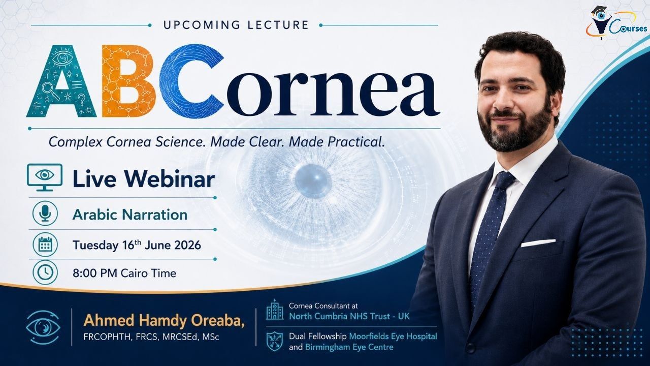
ABCornea
Cornea Basics for Ophthalmology Residents
A practical free webinar on corneal examination, common signs, and clinical fundamentals
Corneal examination can feel challenging early in training. Subtle signs, lesion depth, staining patterns, infiltrates, scars, oedema, vascularisation, and surface disease can all look confusing without a structured approach.
This free webinar is designed to give ophthalmology residents a clear, practical framework for examining the cornea and recognising the common clinical signs encountered in daily practice and exams.
What you will learn
1. A systematic approach to corneal examination
Learn how to assess the cornea from epithelium to endothelium using a logical slit-lamp sequence.
2. How to determine the depth of corneal lesions
Understand practical slit-lamp techniques to localise pathology to the epithelium, stroma, Descemet’s membrane, or endothelium.
3. Common corneal signs every resident must recognise
Identify key signs such as epithelial defects, infiltrates, stromal haze, keratic precipitates, oedema, scarring, vascularisation, thinning, and melt.
4. Fluorescein staining made simple
Learn how to interpret staining patterns in dry eye, epithelial defects, abrasions, ulcers, exposure, neurotrophic disease, ocular surface and contact lens-related disease.
5. Corneal infection vs inflammation
Build confidence in recognising features that suggest microbial keratitis, sterile infiltrates, marginal keratitis, viral keratitis, and immune-mediated disease.
6. Practical clinical pearls for on-call and clinics
Learn what to document, when to worry, when to scrape, when to refer, and how to communicate findings clearly.
Who should attend?
This webinar is ideal for:
- Ophthalmology residents and trainees
- Junior doctors starting ophthalmology
- Optometrists interested in corneal signs
- Exam candidates preparing for FRCOphth, ICO, or FRCS Ophthalmology
- Anyone wanting a structured foundation in corneal assessment
Why attend?
By the end of this session, you will be able to examine the cornea more confidently, describe findings more accurately, and recognise important signs that guide diagnosis and management.
This is not just a theory lecture — it is a practical, image and video based teaching session focused on real clinical examination skills.
Webinar highlights
- Practical slit-lamp examination framework
- Image-based teaching
- Common corneal signs explained simply
- High-yield clinical and exam pearls
- Useful for clinics, on-call, and viva-style exams
- Free registration
Reserve your free place today
Join this practical webinar and build a stronger foundation in corneal examination and diagnosis.
Register now — limited places available.
After Registration, you will be redirected to another page to choose a password. The attendance link will be emailed to you and will be found in your account after logging in
